You are here: Home: NHLU 2 2005 : Steven T Rosen, MD
| Steven T Rosen, MD |
EDITED COMMENTS |
 WHO-EORTC classification for cutaneous lymphomas WHO-EORTC classification for cutaneous lymphomas
Recently, the classification of cutaneous lymphomas has been revised. This was a monumental effort undertaken by the EORTC and WHO, which previously had separate classifications (Willemze 2005).
The cutaneous T-cell and natural killer (NK) cell lymphomas include a host of entities. For the average clinician who may see one case in a lifetime, these may be confusing.
However, almost 90 percent fall into a few categories: (1) mycosis fungoides and its variants; (2) Sézary syndrome, the leukemic form of mycosis fungoides and (3) CD30-positive lymphoproliferative disorders of the skin, including CD30-positive anaplastic lymphomas and its benign counterpart, lymphomatoid papulosis.
The B-cell family of cutaneous lymphomas has three major entities. Primary follicle center lymphomas can be confusing to clinicians, because sometimes the report will say diffuse large cell lymphoma, but diffuse large cell lymphoma of the skin of the follicle center type is treated differently. It can either be observed or treated with local radiation, depending on the clinical circumstances.
The MALT type of lymphoma of the skin is indolent with a 100 percent five year survival rate. It is often treated for cosmetic purposes. The last type, a very unusual entity that is usually seen in elderly women, is a large cell lymphoma that typically tends to be more aggressive and only about half of the patients are alive at five years.
Pathogenesis of cutaneous lymphomas
It has been speculated that viruses cause several cutaneous lymphomas. The human T-cell lymphoma virus type 1 (HTLV-1) has been associated with a form of non-Hodgkin’s lymphoma that often presents in the skin. In fact, the first patient diagnosed with HTLV-1 lymphoma was one I took care of as a fellow.
In the initial report it was called the CR virus, named after the patient who had this unusual presentation of lymphoma. It subsequently was discovered that the CR virus was HTLV-1 and that the patient’s clinical presentation resembled the HTLV-1 type of infections seen in the Kyushu Province of Japan where it’s endemic.
Although this patient had not been to Japan, he had a clinical presentation similar to that seen in patients in the Kyushu Province: Skin lesions, hepatosplenomegaly, a lytic bone lesion with hypercalcemia, brain metastases, opportunistic infections and an expansion of the CD4-positive cells.
Some investigators have provided data suggesting HTLV-1 is present in mycosis fungoides, but others have not verified that. No definitive virus has been associated with the other T-cell processes, but the Epstein-Barr virus has been associated with the NK cell-like T-cell processes involving the skin. One outbreak of B-cell lymphoma in Scotland has been associated with the organism found in Lyme’s disease.
Evaluation of patients with cutaneous lymphomas
The main issues are the clinical presentation (eg, the appearance of the skin) and the biopsy information. The histology and the immunophenotyping and molecular analyses will help direct therapy — especially for patients with B-cell lymphomas in whom you can often have the wrong impression unless you have those data.
Primary cutaneous follicle center lymphomas: Effects of rituximab
This is a fairly common entity. It presents as slightly raised papules or tumors. The biopsy shows a lymphoid infiltrate consistent with lymphoma, and immunophenotyping shows it as a B-cell process that expresses CD20.
Inexperienced clinicians often mistake it for a large cell lymphoma in the skin that should be treated with combination chemotherapy with or without radiation therapy. In reality, you can observe these patients even if they have multiple indolent lesions, unless there’s a cosmetic concern. You can also treat it with local radiation therapy. It’s rare to use chemotherapy as the initial treatment. The five year survival approaches 100 percent.
Rituximab is also effective (Heinzerling 2000, Kennedy 2004). The first patient I treated with rituximab — about five or six years ago — had this entity and presented with two lesions on the face. It was a cosmetic issue and that was the rationale for using rituximab.
The patient also had lesions scattered about his trunk and extremities, and he has been in remission since receiving treatment with rituximab. I’ve treated about a half dozen patients with rituximab, and they have had universal benefit.
Clinical trial of lenalidomide in patients with cutaneous lymphomas
We anticipate beginning accrual to our lenalidomide (Revlimid®) trial within the next four to six weeks. Lenalidomide has been shown to be an effective agent for the treatment of multiple myeloma (Richardson 2002) and, more recently, myelodysplastic syndromes (List 2005). It’s a thalidomide analog and an immune modulator. The mechanism of action is unknown, but speculation exists about its effects on cytokines and angiogenesis.
The drug is well tolerated and is administered orally, which is nice for patients with cutaneous lymphomas who have significant skin disease for whom it can be difficult to use intravenous agents. The main side effects associated with lenalidomide are cytopenias. The patients’ blood counts will be monitored throughout the trial.
Clinical trial of alemtuzumab and rituximab in patients with chronic lymphocytic leukemia
We’re about to initiate a trial of alemtuzumab, targeting the CD52 antigen, and rituximab as the sole up-front therapy in patients with previously untreated chronic lymphocytic leukemia. Patients will receive alemtuzumab subcutaneously three times a week for 16 weeks and rituximab intravenously every two weeks for a total of eight doses. We’re anticipating durable responses without some of the toxicity associated with traditional chemotherapy.
In patients who previously failed traditional chemotherapy and were treated with alemtuzumab and rituximab, we saw responses and acceptable toxicity. The preliminary data indicate no additive toxicity (Nabhan 2004, Faderl 2003). A report from Dr Österborg in Sweden demonstrated that up front, alemtuzumab had results comparable to those of fludarabine and was well tolerated with durable remissions (Österborg 1996).
Currently, none of the therapies we utilize in patients with chronic lymphocytic leukemia are curative. Significant long-term toxicity is associated with fludarabine, when administered alone or with cyclophosphamide, although it is effective. Rituximab combined with chemotherapy appears to be a significant advance. We’re exploring its activity in combination with alemtuzumab as firstline therapy to determine if the combination should be compared in a Phase III trial to other chemotherapy regimens that are considered more standard.
RESORT trial (ECOG-E4402): Maintenance rituximab compared to rituximab re-treatment upon disease progression in patients with low tumor burden indolent lymphomas
A number of investigators who have evaluated rituximab as up-front therapy demonstrated significant activity, durable responses and good tolerability (Colombat 2001, Hainsworth 2002, Ghielmini 2004). One of the critical issues is whether to administer maintenance rituximab or to re-treat at the time of progression. The RESORT trial (ECOG-E4402) will, in part, answer that question.
It will also evaluate some quality-of-life and cost issues, which are critical in the use of agents of this nature. I’ve enrolled only one patient in the study, but I’ve treated a large number of patients with follicular lymphoma with rituximab as the sole agent. I’ve treated the majority of them with maintenance rituximab, after Dr Hainsworth’s study (Hainsworth 2002) demonstrated durability of remission using this approach and after dialogue with patients who preferred a maintenance approach to being re-treated at the time of progression.
When counseling patients about the risks and benefits of maintenance rituximab, I try to provide them with data. Maintenance therapy clearly results in a longer time until the patient requires additional rituximab or other therapy. I also tell them there’s no proof that using maintenance rituximab is advantageous and that we haven’t seen any obvious long-term toxicities associated with maintenance rituximab to preclude my comfort level with using a maintenance approach. ECOG-E4402 will take a number of years to provide a definitive answer about which approach is most prudent.
Select publications
Photo Atlas of Cutaneous Lymphoma
(See audio CD for PowerPoint slides or visit www.NHLUpdate.com.)
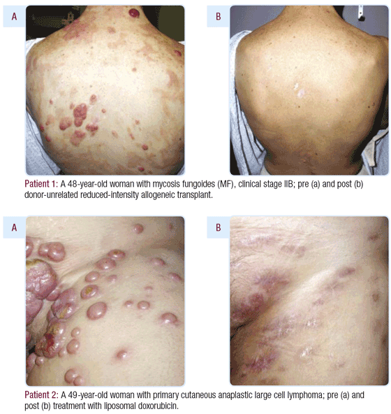
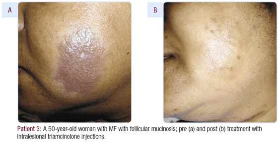
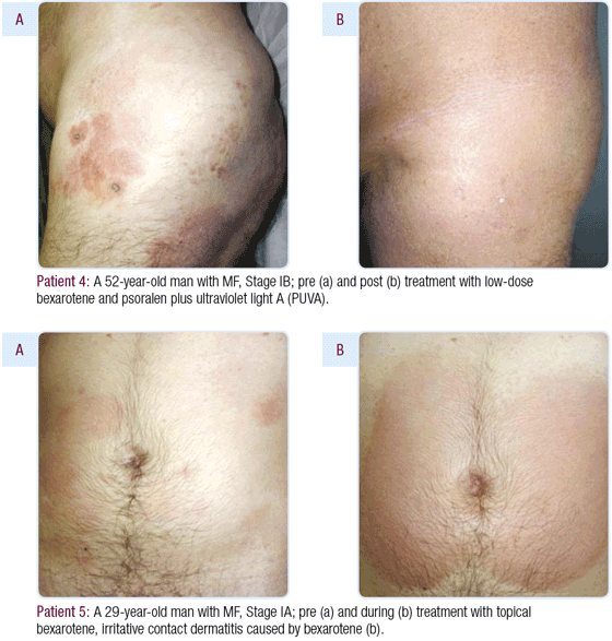
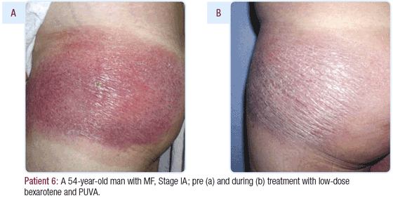
| Dr Rosen is the Director of the Robert H Lurie Comprehensive Cancer Center of Northwestern University in Chicago, Illinois. Photo atlas of cutaneous lymphoma courtesy of Christiane Querfeld, MD, Northwestern University, Robert H Lurie Comprehensive Cancer Center. |
|
